


Select your Market
-
{{ userContent.title }}

{{ userContent.lookingForMoreLabel }}


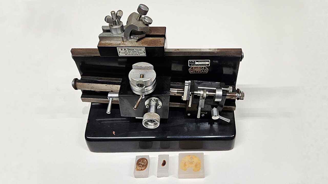
Microtome and histological cuts, 1940s
textblock default title
A microtome is a device used in histological research and diagnostics to produce very thin sections of biological tissue in the micrometer range.
These "histological" sections make it possible to examine the cellular structure of tissue samples in detail. This includes information on tissue composition, cell types, cell distribution and any anomalies. Histology is the science of biological tissue. Staining the sections can also help to differentiate between different cell types and tissue components. As pure cell tissue – even when hardened – is not a substance that can be cut cleanly, it must be embedded in a suitable mass. This is usually kerosene. The water contained in the tissue is replaced by the embedding medium.
The examination of biopsied tissue helps in medicine, for example, in the early detection and classification of tumors, the detection of inflammation or parasitic and bacterial diseases.
Information on the invention of the microtome is not consistent, but it is likely that the Englishman George Adams, Jr. was the one who built such a device in 1770. The term microtome was introduced later. Over the course of time, several variants were constructed, and the shape and type of cutting apparatus was also further developed.
There are several microtomes in the collection here in the archive, which shows the importance of the object for the work at the company. The microtome shown here probably dates from the 1940s.