


Shedding new light onto the structure of ferroelectric hafnium oxide films
Publish Date
02 NOV 2021
Overview
Recently we discovered the presence of a transient crystal structure in hafnium oxide that forms just as it is heated above its crystallization temperature. We were able to “trap” this likely ferroelectric structure before it disappeared to make high performing ferroelectric capacitor.
Over the past decade, scientists have been studying the ferroelectric properties of films based on hafnium oxide. As we’ve described in previous blog posts, hafnium oxide is a widely used material compatible with standard processing techniques in the semiconductor industry. When hafnium oxide adopts specific crystal structures, notably ones that don’t have inversion symmetry, it can become ferroelectric. Ferroelectric materials retain an apparent stored charge (remanent polarization) after an electric field is applied and removed, which is a very useful property for information storage and processing. Ferroelectric devices could create new classes of embedded non-volatile memory or artificial synapses for neuromorphic computing.
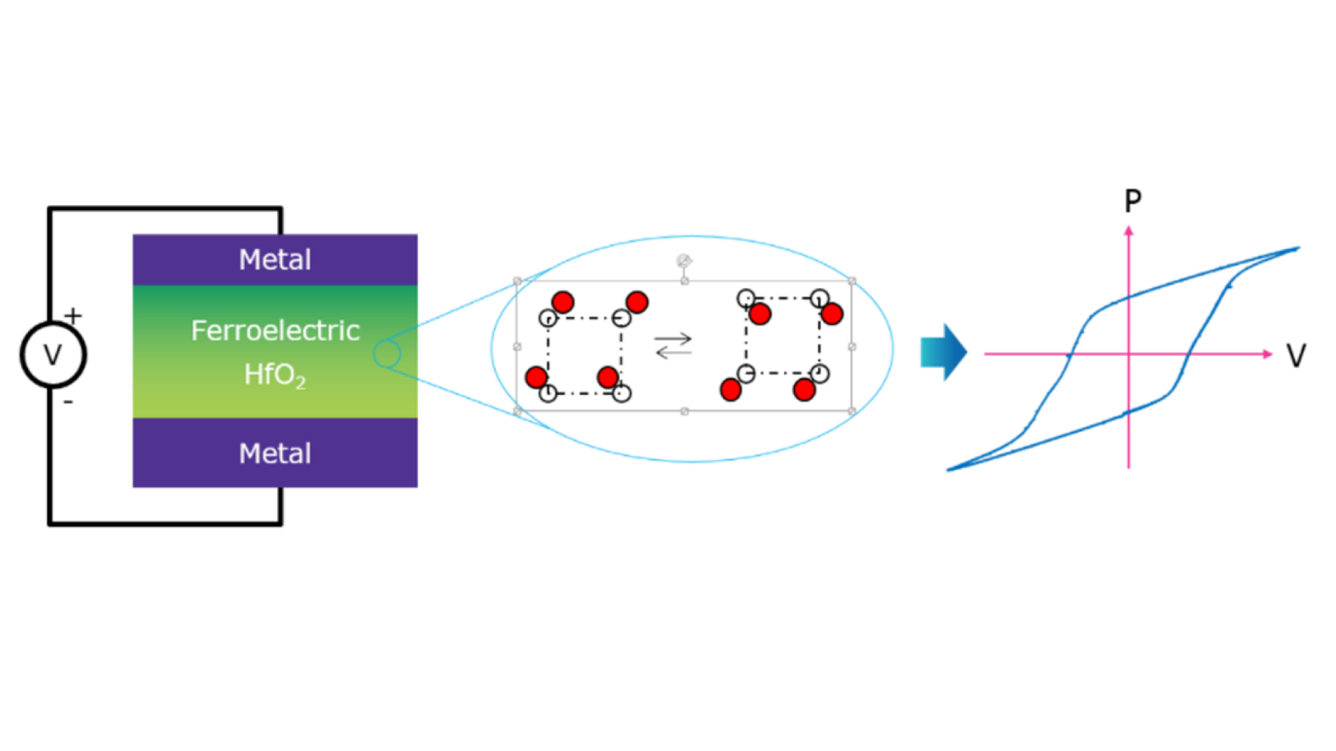
When sandwiched between two conductors, ferroelectric materials form non-linear capacitors. Materials that have no inversion symmetry in the unit cells of their crystal structures can undergo polarization of their lattice. They can thus have an apparent “stored charge” called the “remanent polarization,” Pr
The specific crystal structures needed for ferroelectricity in hafnium oxide aren’t typically the ones that you would observe in crystalline hafnium oxide; normally, the film assumes a monoclinic, non-ferroelectric structure. To stabilize a ferroelectric phase, people have mixed in other materials like zirconium oxide, doped the hafnium oxide with a few atomic percent of another material, like silicon oxide, carefully controlled the thickness and interfaces of the film, and applied specific time-temperature profiles during crystallization. In particular, using rapid thermal annealing equipment or flash lamps seems to be very beneficial for generating a ferroelectric phase in these films. In these types of systems, a sample can be heated to several hundred degrees Celsius within seconds.

Rapid thermal annealers, like this Applied Materials Radiance tool similar to a model used at Intermolecular, uses powerful lamps to rapidly heat a sample. A typical heating profile in an RTP is shown on the right, where a sample can be heated by the lamps to 700°C in about 15 seconds and then cool off slowly once the lamps are turned off.
By performing many experiments, different research groups have attributed the benefits of rapid thermal annealing to the ramp rate, the peak temperature, how long the sample is held at the peak temperature, and the cooling rate. Indeed, all these factors are likely linked together, but a unifying picture was still missing.
Part of the reason that it was difficult to propose a unifying picture is that the changes in the sample were happening on the scale of seconds, but the measurements were being taken on the order of minutes. The gold standard for measuring the crystal structure of films is x-ray diffraction (XRD). X-rays have a wavelength which is on the same scale as the spacing between atoms in a solid. Therefore, when we shine x-rays on a material, the rays hit different planes of atoms in a crystal and scatter at characteristic angles (as defined by Bragg’s law). Each structure forms a specific fingerprint or pattern. On a regular laboratory XRD system, a scan can take 10 to 20 minutes to collect per sample, so any changes to the crystal structure that occur on the scale of seconds would be completely missed.
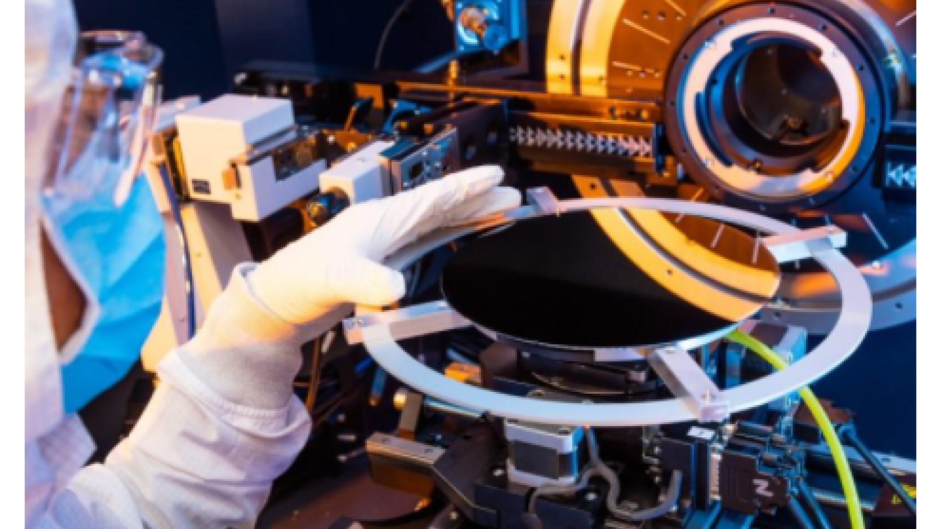
Here, Intermolecular operations team member Hai Huynh loads a wafer on a laboratory x-ray diffraction system. This system uses a point detector, which is the white box mounted on the arm on the left of this photo. To determine the crystal structure of the film on the wafer, the arm is rotated slowly around the circle in the center of the photo to collect the scattered rays. Below, you can see an example of the type of the data that are collected during these measurements. The sample on the top has a tetragonal or orthorhombic crystal phase, which may be ferroelectric, whereas the one on the bottom has a monoclinic crystal phase. Note that the two samples have very different curves, or XRD “patterns.” These patterns take several minutes each to collect, far too slow to capture behavior happening within the first few seconds of crystallization in an RTP.
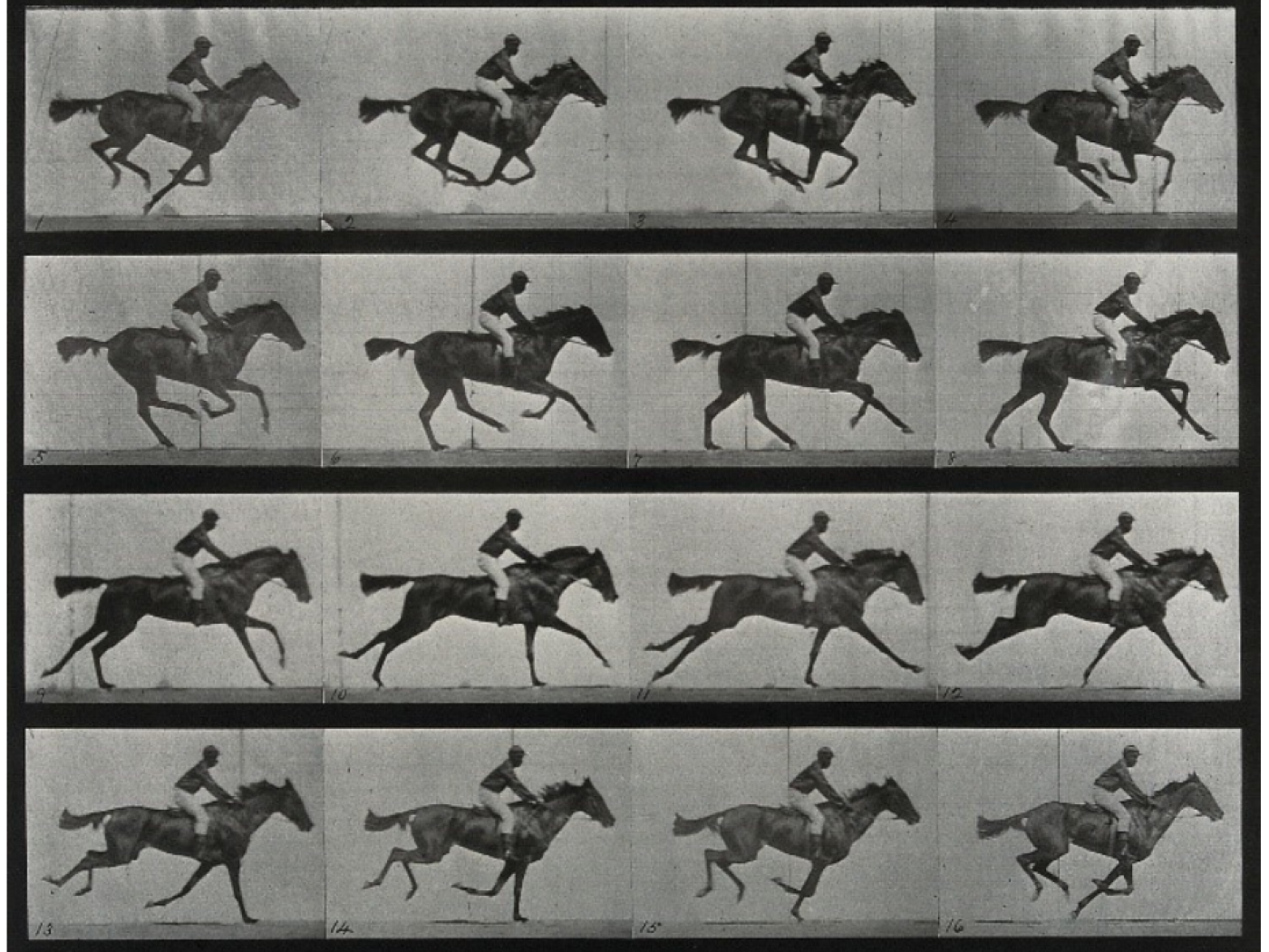
Interestingly, the problem of being unable to collect information quickly enough to understand the dynamics of a system is not a new problem. One example of this problem led to one of the first discoveries made at Stanford University (even before it was a university). Until the late 1800s, most people thought that horses gallop the same way that dogs run, with their limbs splayed out as they jump in the air. However, the actual positions of a horse’s legs when it was in full gallop were too fast for the human eye to clearly see. In fact, no one knew for sure if the horse lifted all four legs off the ground when it was galloping! In the 1870s, Leland Stanford (railroad tycoon and former Governor of California), hired the gifted photographer Eadweard Muybridge to gather the data to resolve this debate once and for all. In 1878, Muybridge was able to create a sequence of photos that clearly showed that a racehorse does in fact lift all four legs in the air, but with the legs tucked below the body. The key technological advance that made this discovery possible was a line of cameras along the racetrack at Stanford’s Stock Farm (now part of Stanford University campus) that sequentially fired to capture the motion of the horse. The cameras were able to do what human eyes could not because they matched the timescale of the measurement to the timescale of the phenomenon being studied.
Now let’s return to the problem of determining why rapid thermal annealing is so beneficial for obtaining ferroelectricity in hafnium oxide. To understand this phenomenon, we needed a way to collect x-ray diffraction patterns much more quickly in order to observe behavior that occurs on the scale of seconds. For starters, instead of a point x-ray detector, we could use a line detector. This would allow us to capture the entire x-ray diffraction pattern in one shot. However, the signal from a standard x-ray source on a thin film of hafnium oxide would produce too few scattered x-rays to detect if we were measuring on this detector at a rate of say one frame per second. To solve this problem, we turned to our friends at SLAC National Lab. Just down the street from the site of Muybridge’s experiment is a large machine that accelerates electrons close to the speed of light. These electrons whiz around a giant ring (about the same size as a football field). As these particles move around through a series of magnetic deflectors, they give off incredibly bright beams of light, including x-rays.
We used a specially designed RTP chamber which we could place between the synchrotron x-rays and a thin area detector (which acts like a line detector). With the ultra-bright x-rays and the new detector, we only needed one second to capture an x-ray diffraction pattern on this system instead of 20 minutes on a standard laboratory system! That means that we could look into what was happening to our hafnium oxide films in situ, while they were being heated up in a rapid thermal annealer.
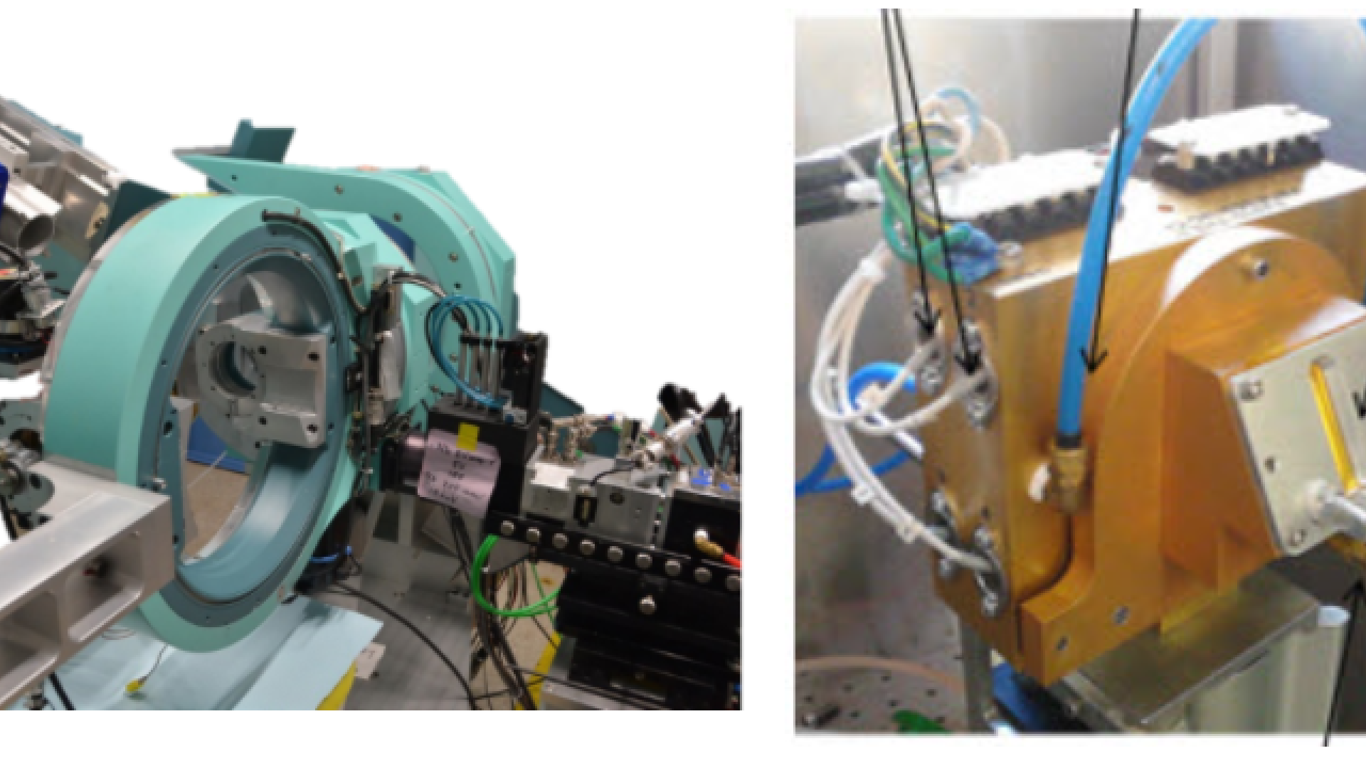
The x-ray diffractometer we used for this study has a line (thin area) detector, the blue box with the silver window at the top left. This enabled us to collect an entire x-ray diffraction pattern in a single shot. The RTP chamber we mounted onto the beamline (on the right) had small windows to allow x-rays to enter and exit, allowing us to do measurements in situ while we heated the sample.
After setting up the system, we started heating up samples of ALD-grown hafnium oxide sandwiched between two titanium nitride conductive layers; this is a typical capacitor stack. What we found was pretty amazing. About 1 second after the film first started to crystalize, the XRD pattern showed a peak characteristic of a ferroelectric phase, which is exactly the phase we were hoping for! However, a few seconds later, this peak started to disappear, and a pair of peaks appear on either side of it. This means that the film was transforming to the monoclinic phase, a more stable but non-ferroelectric structure.
12 seconds worth of XRD patterns taken at one frame per second. This set of patterns shows the crystallization behavior of 7 nm ALD hafnium oxide at a ramp rate of 30°C/s. The first frame shows the amorphous, non-crystalline film. The next two frames show the initial crystallization pattern, which we identified as likely being from a ferroelectric phase. Note the change in the pattern over the next several frames, which indicates a shift from the initial ferroelectric phase to the monoclinic phase.
We repeated these measurements across many samples at different ramp rates, and we noticed that films heated at fast ramp rates retained a higher fraction of ferroelectric phase when the annealing was complete. More than just providing pictures of this transformation, these experiments also gave us the key to understanding how to trap the ferroelectric phase before it disappeared. We developed a kinetic model of the transformation and determined that there was an energetic barrier of over 1 electron volt between the initial ferroelectric phase and the monoclinic phase. A barrier this large explains all the observations we made and that others have made in the literature; it takes time and energy to overcome this barrier, so by limiting either one, we may be able to trap the system in the metastable ferroelectric structure. This could include increasing the ramp rate, decreasing the time at the maximum temperature, or quickly cooling the chamber down after annealing.
We decided to try this by annealing two samples: one had the temperature ramped up slowly (10°C/s), while the other was ramped very fast (50°C/s). We measured the films both using x-ray diffraction and electrical testing, and we determined that the film ramped up quickly had a higher fraction of the ferroelectric phase in the film as well as a higher remanent polarization.
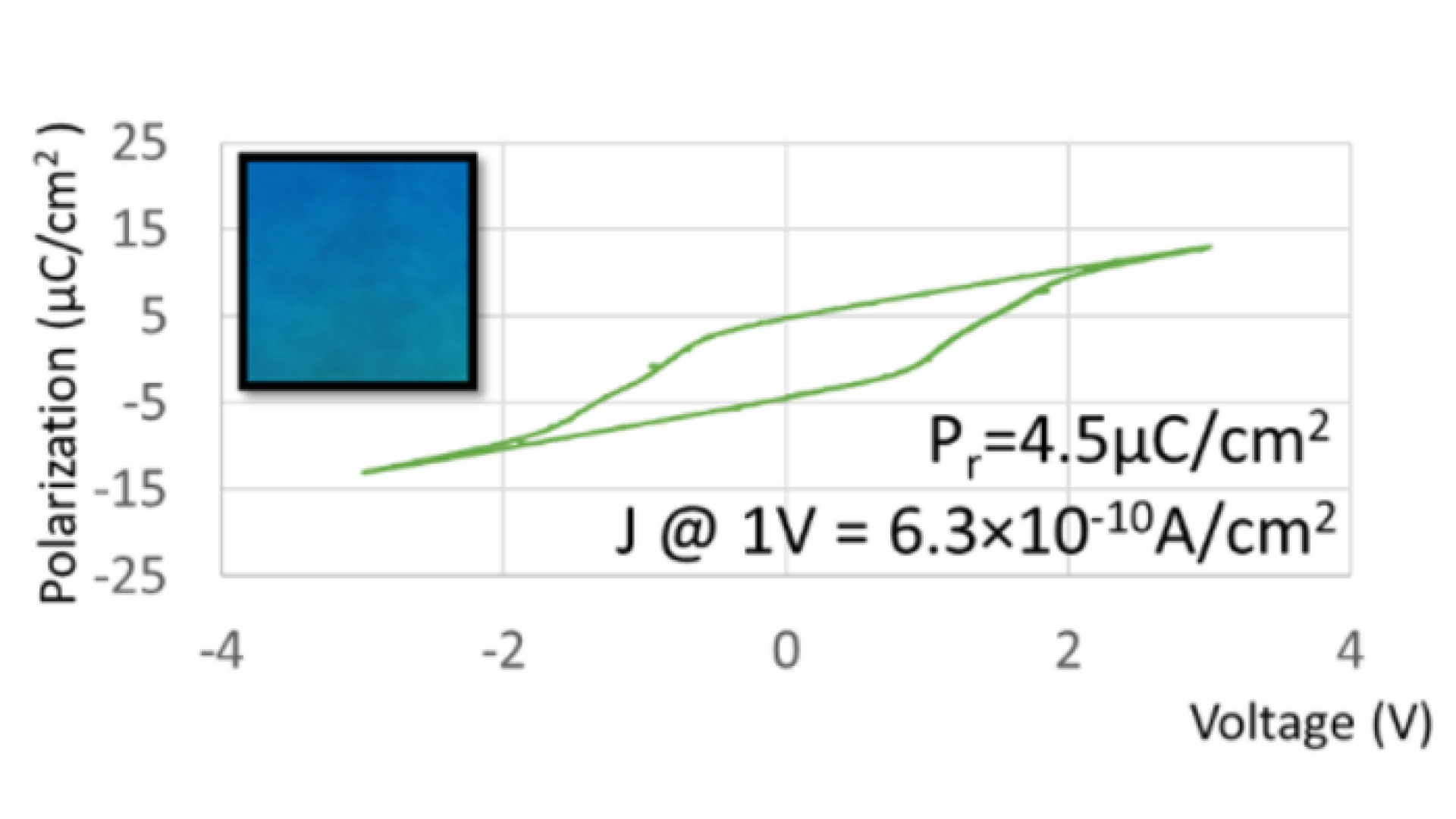
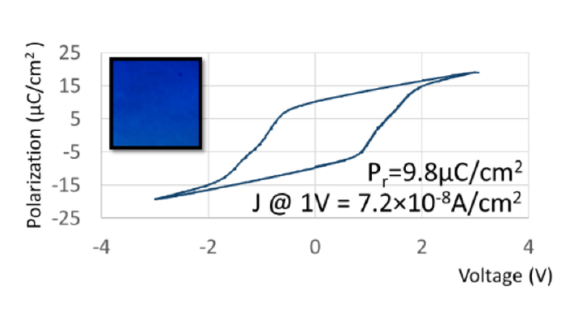
A hafnium oxide film annealed to 700°C with a heating rate of 10°C/sec (left) has a lower remanent polarization than one annealed with a heating rate of 50°C/sec (right). Not only that, but the films actually had different colors – the sample with the higher ferroelectric fraction was very blue, while the one with the lower ferroelectric fraction was more green!
In summary, using synchrotron radiation to very rapidly collect x-ray diffraction data from hafnium oxide capacitors while they were being annealed unveiled hidden behavior that occurs on the timescale of seconds. This collaboration gave us a deep understanding of the kinetics at play in ferroelectric hafnium oxide and a pragmatic approach to making devices with optimized ferroelectric behavior- and opening up new application possibilities.